MID 35 Peptide: Potency, Mechanisms, and Clinical Data
The mid 35 peptide is an experimental myostatin-inhibitory compound currently evaluated in murine models for its localized effects on skeletal muscle protein synthesis and bioactive sphingolipid metabolism. There are no published human clinical trials or pharmacokinetic studies for this compound, categorizing it strictly as a research chemical rather than an approved therapeutic intervention.
01 — What the peptide targets
The mid 35 peptide is a synthetic D-peptide engineered to bind and inhibit myostatin, a primary negative regulator of muscle tissue accumulation. In laboratory HEK293 reporter assays, the compound demonstrated high binding efficiency, establishing a specific potency threshold that differentiates it from earlier macromolecular myostatin inhibitors.
Researchers developed this specific sequence to improve the stability and efficacy of localized myostatin blockade. D-peptides are composed of D-amino acids, which are chiral enantiomers of naturally occurring L-amino acids. This structural modification renders them highly resistant to endogenous proteolytic enzymes, substantially increasing their half-life in a testing medium compared to native peptides. In evaluating cellular assays, researchers observed an IC50 of 0.19 ± 0.05 µM for myostatin, allowing scientists to accurately model the receptor saturation thresholds necessary to blunt myostatin's catabolic signals. In broader evaluations of the Best Peptides for Muscle Growth, researchers categorize MID-35 separately from growth hormone secretagogues due to this direct receptor interference.
02 — Tracking lipid metabolism
Inhibiting myostatin alters bioactive sphingolipid metabolism concurrently with initiating skeletal muscle hypertrophy. This metabolic shift indicates that the peptide influences complex intracellular signaling networks and membrane dynamics, rather than simply acting as a mechanical block on the myostatin receptor pathway.
Sphingolipids like ceramide and sphingosine-1-phosphate function as critical signaling molecules mediating cellular stress, survival, and structural integrity. A controlled murine study observed that administering myostatin inhibitory d-peptides induce skeletal muscle hypertrophy along with alteration of bioactive sphingolipid metabolism. This intersection demonstrates that physiological signaling involves significant lipid profile remodeling within the muscle tissue, complicating the measurement of isolated protein synthesis. The observed shift in lipid profiles emphasizes that true muscle growth requires a fundamental reorganization of the cell membrane and underlying signaling infrastructure.
03 — Regional muscle growth metrics
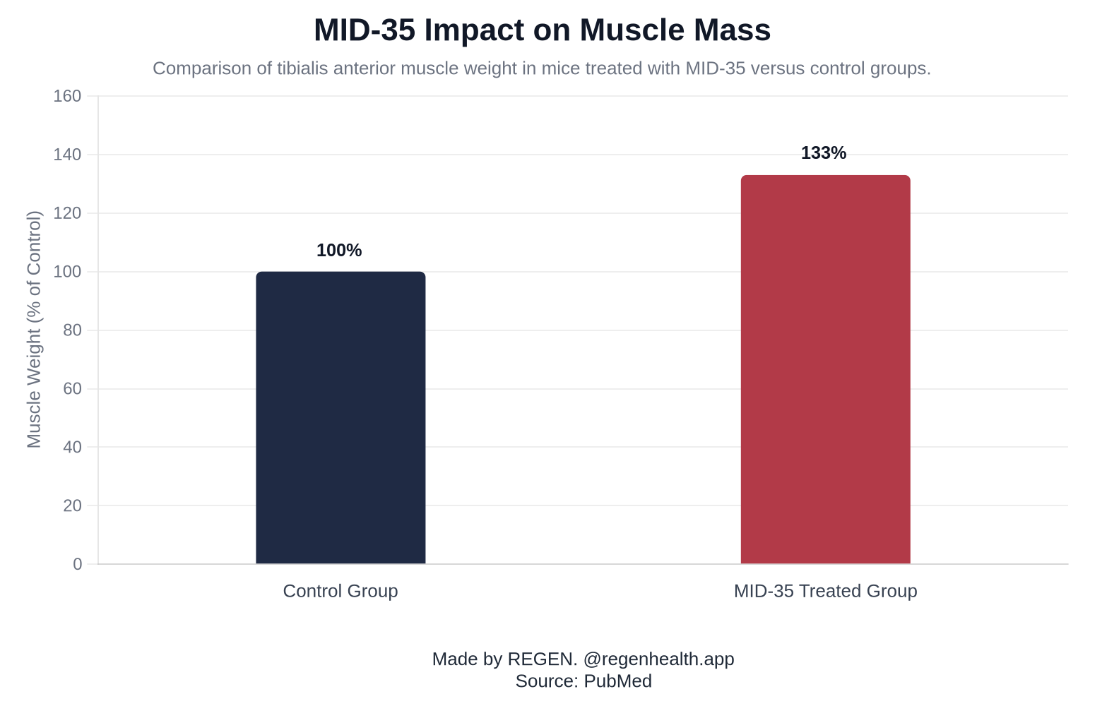
Intramuscular administration significantly increases regional muscle mass in rodent models under laboratory conditions. Data confirm that direct localized injections prompt measurable hypertrophic responses in the targeted skeletal muscle tissues over a four-week evaluation period without immediate systemic dispersion.
The exact study parameters are crucial for contextualizing the hypertrophic response. In a controlled study tracking morphological outcomes, researchers administered an intramuscular injection of 30 nmol MID-35 to mice. By day 28, the treated subjects exhibited a 133% tibialis anterior muscle weight compared to control animals. These metrics quantify the localized hypertrophic potential of the D-peptide under highly controlled, site-specific delivery conditions. However, researchers caution that this magnitude of isolated regional growth in a murine tibialis anterior does not automatically translate to proportional systemic muscle accrual across the entire organism, necessitating further investigation into systemic delivery constraints.
04 — Non-invasive iontophoresis delivery
Because frequent intramuscular injections create localized tissue trauma, researchers test non-invasive delivery methods for myostatin inhibitory peptides. Iontophoresis uses a mild electrical current to drive charged peptide molecules through the skin barrier directly into the underlying muscle tissue.
By leveraging iontophoresis, a study successfully bypassed the epidermal barrier without physical puncturing. This trial focused on increasing skeletal muscle mass in mice by non-invasive intramuscular delivery of myostatin inhibitory peptide by iontophoresis, opening new avenues for repeated-dose studies. This electrical transdermal delivery mechanism maintains the structural integrity of the muscle tissue that would otherwise be inflamed by daily mechanical injections, ensuring that the measured hypertrophic response is solely due to the peptide's biochemical action rather than an inflammatory repair artifact.
05 — The hypertrophic paradox
Inducing rapid muscle mass increases through isolated myostatin inhibition carries the risk of a hypertrophic paradox, where overall tissue weight increases while functional tissue quality and force generation decrease. Monitoring actual physiological signaling requires tracking specific serum biomarkers.
Simply increasing the physical cross-sectional area of a muscle fiber does not guarantee an equivalent increase in contractile force. In laboratory environments exploring this intersection, researchers track baseline and longitudinal Creatine Kinase alongside myostatin serum levels to monitor metabolic distress. If skeletal muscle hypertrophies without commensurate connective tissue and neurological adaptation, the resulting mass may be structurally compromised. Protocols investigating complete tissue remodeling often examine parallel compounds, such as BPC-157, when studying connective tissue integrity alongside muscle growth. The mid 35 peptide is not FDA-approved for human use; sold for research purposes only.
06 — Clinical status and human data
There are no published human clinical trials or pharmacokinetic studies for MID-35, limiting the understanding of its safety, systemic distribution, and biological half-life. It remains entirely restricted to preclinical testing and in vitro laboratory investigation.
Because of this complete absence of clinical data, researchers cannot determine appropriate human clearance rates or long-term organ toxicity. The exact metabolic fate of the peptide in human circulation is unknown. Those researching muscle signaling pathways in human subjects often rely on tracking baseline blood chemistry to identify abnormally high baseline myostatin levels before considering theoretical interventions. Compounds with established human pharmacokinetic profiles, such as Tesamorelin, are reviewed in distinct discussions of body composition therapies, though they operate via entirely different endocrine pathways. The lack of clinical phase testing means that any theoretical application of MID-35 relies purely on extrapolating murine data, a practice with a historically high failure rate.
FAQ
Is it safe to take myostatin inhibitors?
Myostatin inhibitors remain experimental compounds with largely unknown long-term safety profiles in humans. Without extensive clinical trials, the risks include potential structural weaknesses in connective tissues, altered metabolic signaling, and unforeseen systemic side effects.
What is the strongest HGH peptide?
The strength of an HGH-related peptide depends heavily on the targeted metabolic or morphological outcome rather than a single absolute metric. Compounds like Ipamorelin and Tesamorelin are frequently cited in research for their specific secretagogue effects, but none are universally classified as the single strongest.
Is follistatin legal to buy?
Follistatin is not FDA-approved for human consumption and cannot be legally sold as a medication or dietary supplement. It is legally available exclusively as a research chemical intended strictly for in vitro or animal laboratory testing.
What limits muscle growth?
Muscle growth is primarily limited by genetic and biochemical factors, most notably the continuous presence of myostatin. This protein signals the body to halt excessive muscle tissue accumulation, serving as a biological governor to prevent disproportionate metabolic energy demands.